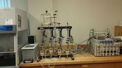
 Figure 1. Ex vivo muscle function apparatus
Materials and Methods:
All procedures were done according to the Institutional Animal and Use Committee. Ten week old Sprague- Dawley rats were randomly categorized into four groups (N=3). These experimental groups were consisted of: Krebs buffer (K-K) our control, Creatine buffer (C-K), Doxorubicin buffer (Dox-K), or Creatine and Doxorubicin buffer (C-D). Four buffers were made and each buffer contained a simple Krebs buffer. Krebs buffer is composed of: 12.252 grams of Glucose, 28.048 grams of Sodium Chloride, 1.468 grams of Calcium Chloride, 1.76 grams of Potassium Chloride, 8.4 grams of Sodium Bicarbonate, and .96 grams of Magnesium Chloride. In the Creatine buffer, a Krebs buffer was compounded with 25 mM of Creatine. In the Dox buffer, a Krebs buffer was affixed with 25 μM of Dox.
The extensor digitorum longus (EDL) and soleus (SOL) were extracted from the left hind limb and stabilized for five minutes in Krebs buffer. After stabilization, sutures with micro spring clips (Fig. 2) were attached to the distal and proximal tendons of each muscle, and then submerged in an organ chamber (Fig. 3) filled with Krebs buffer. Muscle function was then analyzed, by using an ex vivo muscle function apparatus (Fig. 1). The proximal end of the muscle was attached to an isometric force transducer; the distal end was attached to a stationary glass hook (Fig. 3). After attaching the muscles to the force transducer, the maximal force determination was made by adjusting both voltage and tension until maximal twitch force was obtained.
Next, the muscles were changed and treated with the corresponding buffers (C-K, D-K, or C-D). The muscles were stimulated during the incubation period, using platinum field stimulating electrodes for 200 milliseconds at 100 Hz once every five minutes for 30 minutes at the same voltage achieved during the maximal force determinations. The Creatine and Dox buffer was a set of each buffer; the creatine buffer and then the Dox buffer one after another. After the initial incubation period, fresh Krebs buffer was added and the muscles were conducted to a 100 seconds fatigue protocol during which muscle force was recorded every 10 seconds for 100 seconds via Lab Chart software (ADI instruments, Colorado springs, Colorado) using the same voltage as before. After the incubation process, the tissues were frozen in liquid nitrogen. Then, the muscles were homogenized (Fig. 4) in a RITA-Lysis buffer and analyzed for MDA (Malonaldehyde) content.
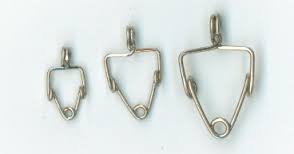
Figure 2. Micro spring clips
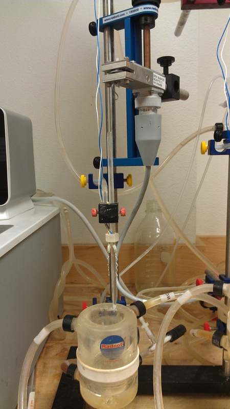
Figure 3. Isometric force transducer on top, stationary glass hook, and organ chamber on the bottom.
|
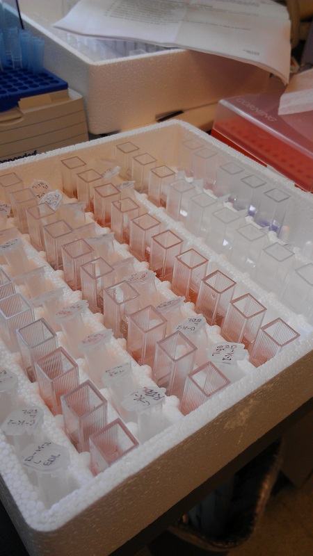
Figure 4. Homogenized muscles
|
